

Abnormal uterine Bleeding
AUB
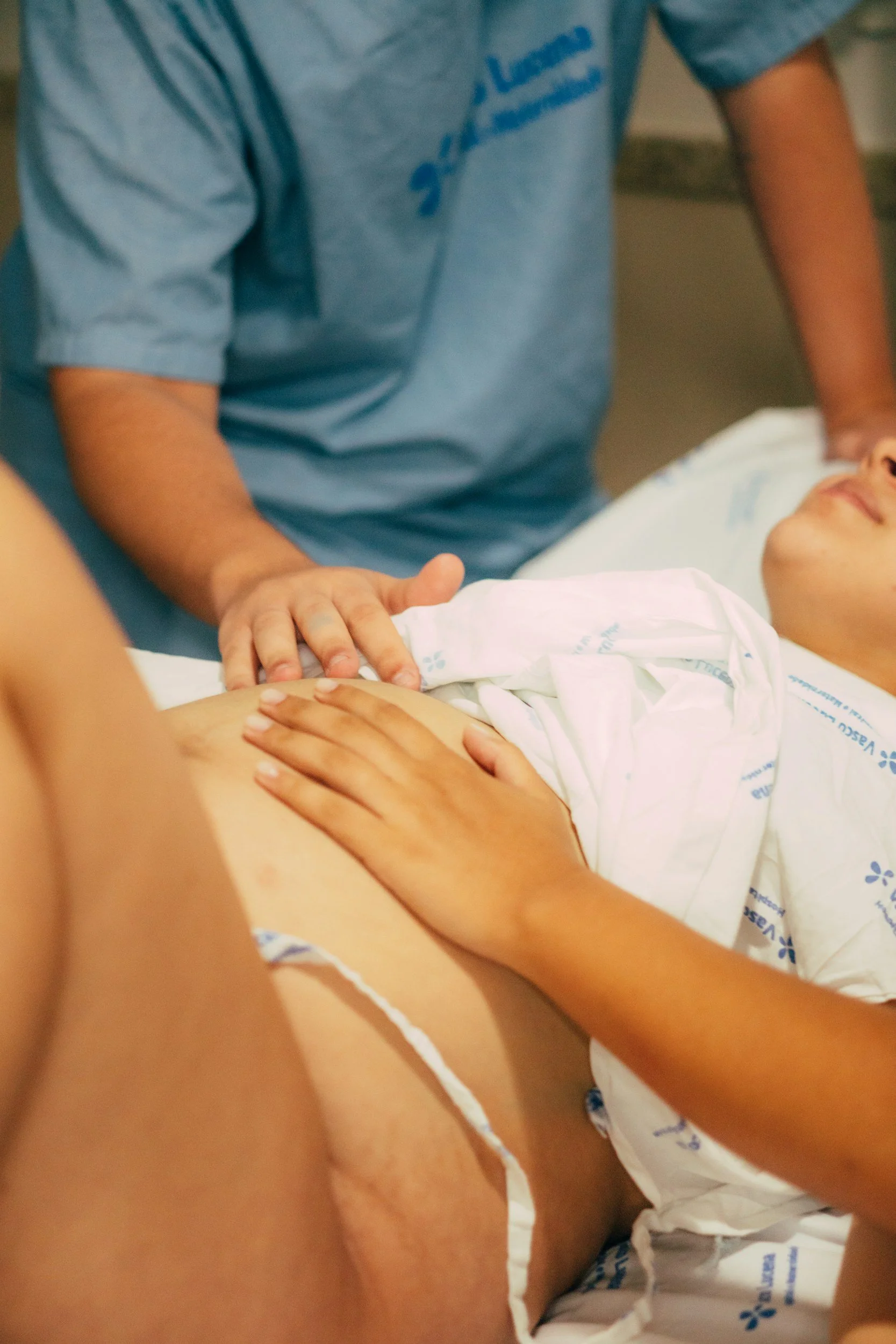
Normal Menstruation
Normal Menstruation
Menstruation is a natural, hormone-driven process in which the lining of the uterus (endometrium) is shed each month when pregnancy does not occur. This cycle is regulated mainly by the hormones oestrogen and progesterone.
A typical menstrual cycle lasts around 28 days, (24-38 days) with bleeding for 4–5 days (2-7 days) with a blood loss of 5–80 mL.

What Happens During a Period
The upper layer of the uterine lining breaks down and is shed
Blood vessels constrict and then reopen, contributing to bleeding
Inflammatory and repair processes are activated
The uterus contracts (often felt as cramps) to help expel menstrual tissue


Mechanisms That Stop Menstrual Bleeding
The body uses several coordinated processes to stop menstrual bleeding:
Uterine contractions: Help compress blood vessels and reduce blood flow
Vasoconstriction: Narrowing of endometrial blood vessels limits bleeding
Endometrial repair: Rapid healing and regeneration of the uterine lining restore normal surface integrity
These mechanisms work together to ensure bleeding remains controlled and self-limited.

How the Cycle Works
Build-up phase (proliferative phase):
Oestrogen stimulates the uterine lining to thicken and prepare for possible pregnancy.
Post-ovulation (secretory phase):
Progesterone stabilises and matures the lining, making it suitable for embryo implantation.
Menstruation:
If pregnancy does not occur, hormone levels fall—especially progesterone—triggering shedding of the lining

Repair and Regeneration
After shedding, the lining rapidly repairs and regenerates from deeper layers, preparing for the next cycle.
Normal menstruation depends on a delicate balance of hormonal, vascular, and immune processes. Disruption of any of these can lead to abnormal bleeding patterns.

Abnormal Uterine Bleeding

Any deviation from the above parameters is considered Abnormal uterine bleeding (AUB). The latter refers to any variation in menstrual cycle frequency, regularity, duration, or volume in non-pregnant women of reproductive age. Up to one-third of women experience AUB, particularly around menarche and perimenopause.
Assessment & Management
Evaluation includes a detailed history, examination, and appropriate investigations (blood tests, imaging, or endometrial sampling). Treatment is tailored to the underlying cause, severity, and patient preferences, with the aim of controlling bleeding and improving quality of life.

Classification
Modern classification (FIGO) improves clarity and includes:
Descriptive terms:
Heavy menstrual bleeding (HMB)
Intermenstrual bleeding
Breakthrough bleeding (on hormonal therapy)
Causes (PALM-COEIN):
Structural: Polyp, adenomyosis, fibroids (leiomyoma), malignancy/hyperplasia
Non-structural: Coagulopathy, ovulatory dysfunction, endometrial, iatrogenic, other
Types
Acute AUB: Sudden, heavy bleeding requiring urgent management
Chronic AUB: Irregular bleeding present for most of the past 6 months
Classification of AUB
PALM-COEIN
The FIGO PALM-COEIN system categorises causes of abnormal uterine bleeding into structural and non-structural groups to guide diagnosis and management.
Structural Causes (PALM)
Polyp (P): Benign growths that may cause intermenstrual bleeding
Adenomyosis (A): Endometrial tissue within the uterine muscle, causing heavy, painful periods
Leiomyoma (L): Fibroids that may lead to heavy or prolonged bleeding
Malignancy/Hyperplasia (M): Abnormal or cancerous changes of the endometrium, often with irregular bleeding


AUB is often multifactorial, requiring a tailored, patient-specific approach combining clinical assessment, imaging, and laboratory evaluation.
Abnormal uterine bleeding (AUB) is common among reproductive-aged women, with an estimated prevalence of 3% to 30%, increasing to 35% or more when irregular and intermenstrual bleeding are included.
Rates are higher around menarche and perimenopause. Heavy menstrual bleeding (HMB) alone affects up to 27% of women in Europe, with some global estimates exceeding 50%.
True prevalence is likely underestimated, as many women do not seek medical care and symptom reporting can be both subjective and variable.
Non-Structural Causes (COEIN)
Coagulopathy (C): Bleeding disorders (e.g. von Willebrand disease)
Ovulatory dysfunction (O): Irregular ovulation (e.g. PCOS, thyroid disorders)
Endometrial (E): Disorders affecting the lining’s ability to control bleeding
Iatrogenic (I): Medication- or treatment-related (e.g. hormonal therapy, anticoagulants)
Not otherwise classified (N): Less common causes (e.g. vascular abnormalities)
Why Does Abnormal Uterine Bleeding Happen?
During a normal menstrual cycle, the build-up and shedding of the uterine lining are tightly regulated by hormones in a coordinated manner. When this hormonal balance is disrupted, bleeding may become heavier, irregular, or prolonged.
AUB can also occur when the body’s natural mechanisms that control bleeding—particularly uterine contractions and blood vessel constriction—are impaired.
In many cases, more than one factor is involved. Identifying the underlying cause is key to effective, personalised treatment.


Common causes include:
Hormonal imbalance: Irregular ovulation can lead to excessive build-up of the lining and unpredictable shedding
Endometrial (lining) dysfunction: The lining may not regulate bleeding or repair effectively
Structural conditions: Fibroids or adenomyosis can affect uterine contractions and blood flow
Conditions that increase pelvic or uterine congestion:
Pelvic congestion syndrome (enlarged pelvic veins)
Fibroids or adenomyosis increasing uterine blood supply
Chronic inflammation (e.g. endometriosis or pelvic inflammatory disease)
Bleeding (clotting) disorders:
Conditions such as von Willebrand disease or other clotting abnormalities can lead to heavier or prolonged bleeding due to impaired blood clotting
Assessment of Abnormal Uterine Bleeding (AUB)
A thorough clinical assessment is essential to identify the cause and evaluate the impact of abnormal uterine bleeding.
Also, a structured, patient-centred approach ensures accurate diagnosis and supports a tailored management plan.
Initial Assessment
Assess hemodynamic stability first in cases of heavy or acute bleeding
Stabilise the patient if required before further evaluation
Clinical History
A detailed history typically includes the following:
Menstrual history
Age at first period (menarche)
Last menstrual period
Cycle pattern: frequency, regularity, duration, and flow
Indicators of heavy bleeding (e.g. frequent pad/tampon changes, clots, flooding, night changes, impact on daily life)
Intermenstrual or postcoital bleeding

Associated symptoms
Pelvic pain
Vaginal discharge
Bowel or bladder symptoms
Symptoms of anaemia (fatigue, dizziness)
Features of hormonal or endocrine disorders


Reproductive and sexual history
Previous pregnancies and mode of delivery
Fertility wishes or subfertility
Current contraception
History of sexually transmitted infections
Cervical screening (smear) history

Medical and social history
Current medications
Personal or family history of bleeding disorders, endocrine conditions, or malignancy
Lifestyle factors (smoking, alcohol, drugs)
Impact on quality of life
Physical Examination in AUB
A focused physical examination helps identify the cause and assess severity.
This structured examination supports accurate diagnosis and guides further investigations and management.
Initial Assessment
Check for hemodynamic instability
Low blood pressure
Rapid pulse)

Endocrine Features
Thyroid examination (enlargement or tenderness)
Signs of hormonal imbalance, such as:
Excess hair growth or acne (suggesting androgen excess)
Features of Cushing’s syndrome (e.g. central weight gain, striae)

General Examination
Vital signs, including blood pressure and BMI
Signs of anaemia (pallor) or bleeding disorders (bruising, petechiae)
In adolescents, assessment of pubertal development

Abdominal and Pelvic Examination
Abdominal palpation for masses
Pelvic examination (speculum and bimanual) where appropriate
Cervical screening and tests for infections if indicated
Evaluation of Abnormal Uterine Bleeding (AUB)
Assessment of AUB involves a combination of blood tests, imaging, and sometimes endometrial sampling to identify the cause and guide treatment.
A structured evaluation helps distinguish between causes and ensures tailored, effective management.
Imaging
Ultrasound (transvaginal or abdominal) – first-line to assess structural causes (e.g. fibroids, polyps, adenomyosis)
Sonohysterography – improves detection of abnormalities داخل the uterine cavity
MRI – reserved for complex or unclear cases

Laboratory Tests
Pregnancy test (essential in all reproductive-age women)
Full blood count (CBC) to check for anaemia
Additional tests (if indicated):
Thyroid function – if symptoms suggest thyroid disease
Hormonal tests – if endocrine causes are suspected
Bleeding disorder screen – especially with heavy or long-standing bleeding
Ferritin – to assess iron stores
Blood grouping/crossmatch – in severe bleeding cases

Endometrial Assessment
Endometrial biopsy recommended:
Age ≥45 years
Persistent or unexplained bleeding
Risk factors for endometrial cancer
Failed medical treatment
Hysteroscopy may be needed if biopsy is inconclusive or symptoms persist

.Treatment of Abnormal Uterine Bleeding (AUB)
Management depends on the cause, severity of bleeding, fertility wishes, and overall health. Treatment is recommended when bleeding causes anaemia or significantly affects quality of life.
A personalised, stepwise approach ensures effective symptom control while aligning with the patient’s preferences and reproductive goals.
Acute (Emergency) Management
For severe or unstable bleeding:
Hospital admission may be required
Medical treatment:
High-dose hormonal therapy (oestrogen, combined pill, or progestins)
Tranexamic acid to reduce bleeding
Supportive care:
IV fluids, antiemetics, iron
Procedures (if needed):
Uterine tamponade (balloon/catheter)
Rarely surgery (e.g. curettage, embolisation, hysterectomy)

Long-Term Management
Ongoing hormonal therapy to regulate cycles
Iron supplementation if anaemia is present
In selected cases: GnRH analogues (with add-back therapy)


Surgical Options
Considered if medical treatment fails or is not suitable:
Hysteroscopy (polyp removal)
Myomectomy (fibroid removal)
Endometrial ablation (not suitable if future fertility desired)
Uterine artery embolisation
Hysterectomy (definitive treatment)
Non-Emergency (Routine) Treatment
Hormonal options
Hormonal coil (Mirena/LNG-IUD) – most effective for reducing bleeding
Combined oral contraceptive pill – regulates cycles and reduces flow
Progestin-only treatments – oral or injectable
Non-hormonal options
Tranexamic acid – reduces blood loss during periods
NSAIDs (e.g. ibuprofen) – reduce bleeding and pain
Management Based on Cause (PALM-COEIN)
Treatment of abnormal uterine bleeding is tailored to the underlying cause.
Management is individualised, balancing effectiveness, side effects, fertility wishes, and patient preference.
Imaging
Ultrasound (transvaginal or abdominal) – first-line to assess structural causes (e.g. fibroids, polyps, adenomyosis)
Sonohysterography – improves detection of abnormalities داخل the uterine cavity
MRI – reserved for complex or unclear cases
Structural Causes (PALM)
Polyps (P)
Surgical removal (hysteroscopic resection)
Adenomyosis (A)
Definitive: hysterectomy
Selected cases: conservative surgery or hormonal therapy
Fibroids / Leiomyomas (L)
Medical: hormonal coil, GnRH analogues, progestins, tranexamic acid, NSAIDs
Surgical: myomectomy, uterine artery embolisation, endometrial ablation, or hysterectomy
Malignancy / Hyperplasia (M)
Primarily surgical management ± additional therapies (e.g. hormonal or radiotherapy)
Non-surgical options (e.g. high-dose progestins) in selected cases


Non-Structural Causes (COEIN)
Coagulopathies (C)
Tranexamic acid or desmopressin (DDAVP)
Ovulatory Dysfunction (O)
Treat underlying cause (e.g. PCOS, thyroid disorders)
Lifestyle changes and hormonal regulation
Endometrial Causes (E)
Managed symptomatically (no specific targeted therapy)
Iatrogenic (I)
Adjust or change causative medication
Consider alternative contraception if needed
Not Otherwise Classified (N)
Treat underlying condition (e.g. antibiotics for infection, embolisation for vascular issues)